
We use a variety of techniques to study the function of proteins inside and outside cells as well as cell behavior inside and outside organ tissues.
Traction Force Microscopy
Traction Force Microscopy (TFM) measures the contractile force of cultured neonatal mouse/rat vnetricular cardiomyocytes (NMVMs/NRVMs) as well as human-induced pluripotent stem cell-derived cardiomyocytes (hiPSC-CMs). This techniques measures the contractile force in single cardiomyocytes in each beat by measuring the displacement of beads embedded in the cellular substrate with known mechnaical stiffness.
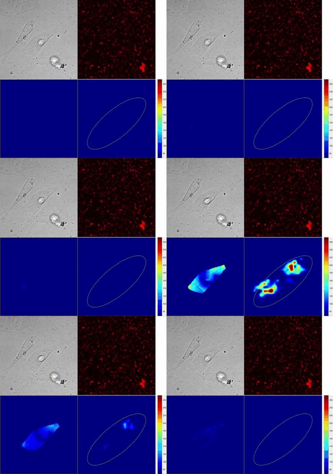
Fluorescence recovery after photobleaching
Fluorescence recovery after photobleaching (FRAP) measures the dynamics of proteins inside cells by measuring the rate of recovery of light from fluorescently labeled proteins that exchange at a particular cellular compartment.
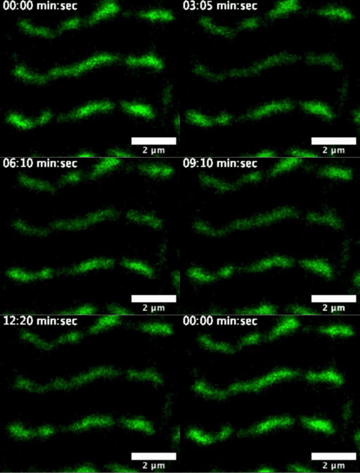
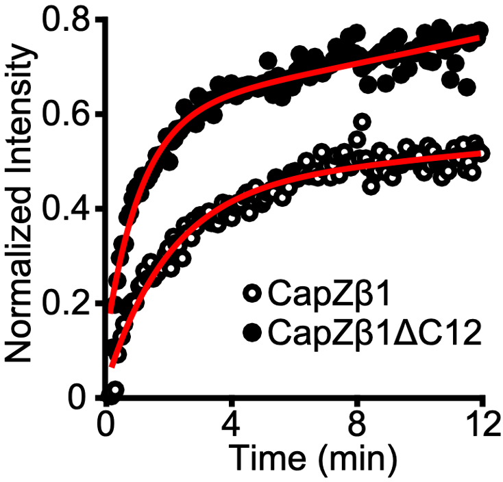
Solís and Russell (2019) J. Gen. Phys. 151, 660.
Förster Resonance Energy Transfer
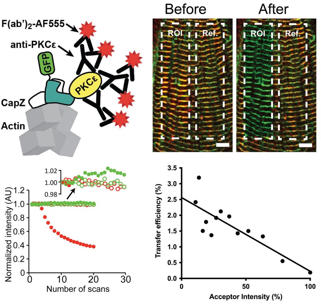
Förster Resonance Energy Transfer (FRET) is a powerful techniques that allows to quantify protein-protein an ligand interactions within cells in the using fluorescence light. The working principle of the techniques relies on two different fluorescent dye molecules tagged covalently or non-covalently to protein domains or ligands. When the labeled proteins or ligands come to a proximity range of >10 nm, a change in the light being emitted is an indicator of such close-proximity interactions taking place.
Cell culture straining
With every heart beat our muscles undergo cycles of shortening during ventricular contraction (i.e. blood is squeezed out from the ventricles) and stretching during ventricular relaxation (i.e. blood fills up the ventricles). We can recapitulate this ventricular stretching in cardiomyocyte cultures by plating primary or stem cell-derived cardiomyocytes in flexible membranes and applying cyclical intervals of stretching. After applying a particular stretching protocol we can use the membranes for immunochemistry imaging, Western blots, RNA sequencing, or mass spectrometry.
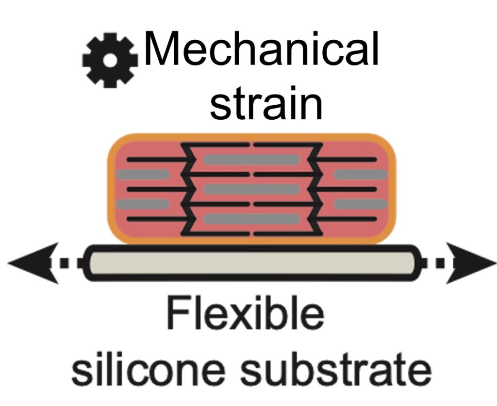
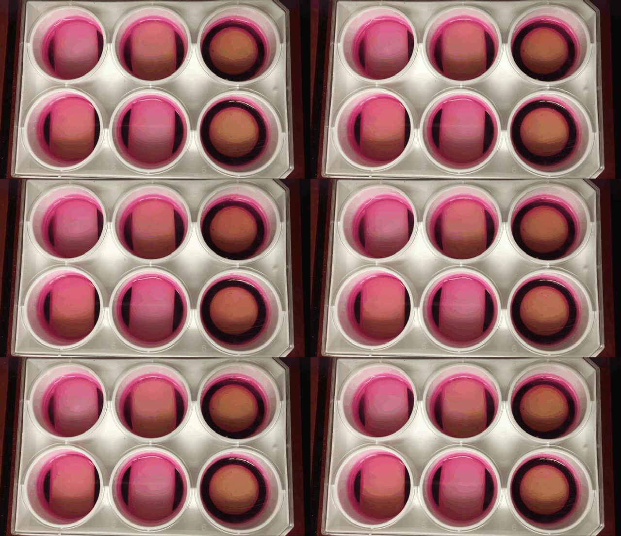
Left: diagram of a cadiomyocyte plated on a flexible silicone membrane. Right: Cell culture strain unit at work. Credit, Christopher Solis
Targeted Mass Spectrometry
Targeted Mass Spectrometry is a powerful technique to study the post-transaltional composition of single proteins. While mass spectrometry can swath across the whole collection of proteins found in a sample and attempt to match these the detected ions against a data base, targeted mass spectrometry focuses on a single protein of interest. This technique focuses on quantifying specific modifications in a particular protein across different samples and treatments at the expense of ignoring any other proteins present in a sample.
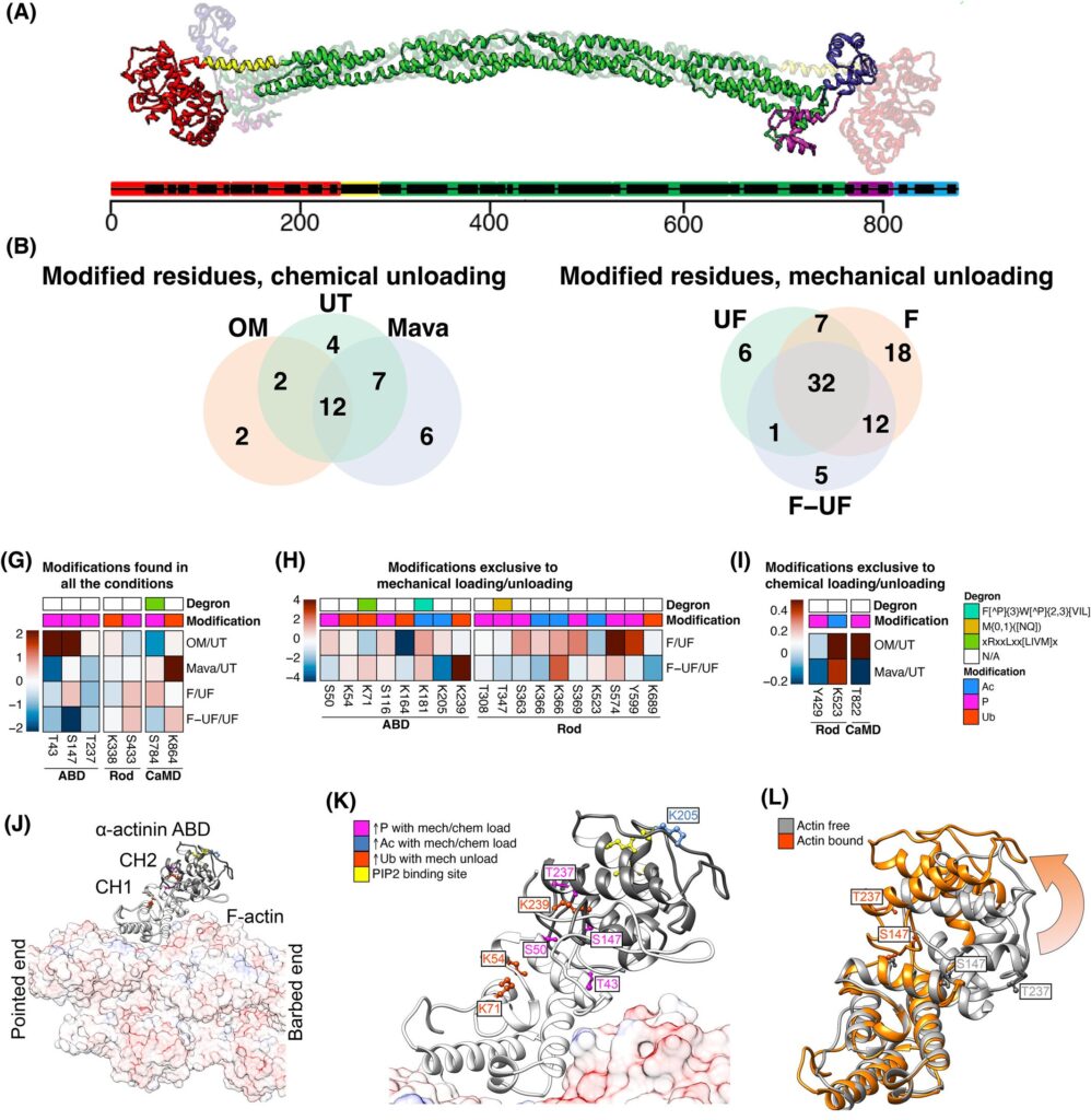
Solís et al. (2023) FEBS J.
In vitro motility assays
The in vitro motility assay was pioneered by the Spudich Lab in the mid 80s-90s. This is a biochemical assay that allows scientists to quantify the level of activity of contractile proteins found in any cell. At minimum, this assay requires myosin to be fixed to a glass surface to drive the chemical conversion of ATP into mechanical propulsion of actin filaments freely gliding on top. In the process videos of fluorecent actin filaments are recorded by fluorescence microscopy. The gliding velocity is a direct measure of biochemical activity from myosin. Variation of these assays may include actin bidnign proteins like troponin and tropomyosin to provide calcium-dependent sliding of actin filaments as well as the inclusion of other actin binding proteins on the surface along with myosin to provide viscoelastic dragging forces to study protein-protein interactions.
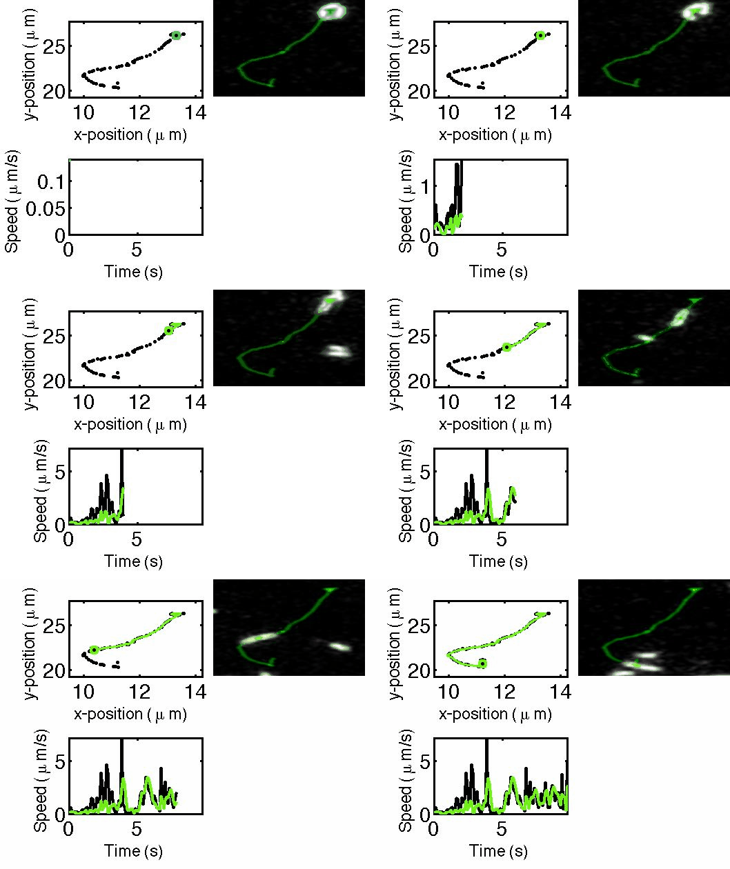
Solís 2025. All rights reserved.
